Low-Energy Electron Holography Imaging of Conformational Variability of Single Antibody Molecules
Single molecule imaging of monoclonal antibodies provides insights into the structural flexibility of this important class of proteins.
Proteins are large and complex macromolecules, which perform a wide variety of biological functions. Their biological functionality is often connected to structural variations, such as stoichiometry, flexible subunits, or changes in folding. Mapping this conformational variability at a sub-molecular scale is thus of high relevance for understanding protein function and behaviour. While the principal methods employed for protein structure determination, such as X‑ray crystallography, NMR spectroscopy and cryogenic electron microscopy, are capable of sub‑molecular and even atomic resolution, they all rely on ensemble averaging, which effectively limits their ability of accessing protein conformational spaces and thus the study of inherently flexible proteins, such as antibodies.
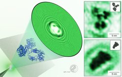
; IgG molecules imaged with our LEEH microscope in its characteristic Y-shaped structure (top right) and showing a gas-phase-related conformation (bottom right) along with the respective structural models (insets).")
Our approach, based on low-energy electron holography (LEEH) microscopy imaging combined with native electrospray ion beam deposition (ES-IBD), allows the imaging of proteins on the single-molecule level without the need for averaging, and thus enables the exploration of the complete conformational space of flexible Immunoglobulin antibody (IgG) molecules. LEEH allows the imaging of individual molecules with high contrast and negligible radiation damage, while native ES-IBD offers the possibility to deposit biologically relevant molecular states on a surface. The combination of these methods thus enables the non‑destructive imaging of individual, native‑like protein molecules.
Herceptin antibodies were gently transferred from a native solution environment into ultrahigh vacuum via a gas phase transition and then landed on a single layer graphene substrate with a range of kinetic energies. A low-energy electron beam (ca. 100 eV) was used to image the deposited molecules in an in-line holography scheme. The hologram, emerging from the interference between the electron waves scattered by the object and the unaltered transmitted wave, is visualized on the phosphorus screen of a microchannel plate detector and captured by a camera. The images of the individual molecules are reconstructed from the hologram by a one-step propagation-based algorithm.
Our LEEH microscope can image IgG molecules with a resolution of up to 5Å, allowing for the distinction of the different antibody subunits as well as other structural features such as the hinge region. Different conformations and subunit orientations can be distinguished and mapped to the crystallographic model. To analyze the large variety of different conformations and adsorption geometries, more than 2000 individual molecules were investigated. This allowed us to identify the role played by the sample preparation process in shaping the molecular conformations observed on the surface. Structures reminiscent of the gas phase conformations adopted by the flexible IgG molecules during the electrospray ion beam deposition process were identified, providing insights for applications of structural biology in the gas phase. Additionally, the fine control of the landing energy enabled us to discern conformational changes induced by the molecule-surface interaction during the deposition process. Our single molecule imaging technique can thus provide valuable information especially when applied to flexible protein structures.


